Home
 White PapersWhite Papers
The University of California, San Francisco's EmbryoScope adoption improved IVF efficiency, saving 7 hours daily and boosting embryo evaluation precision with AI-ready tech.


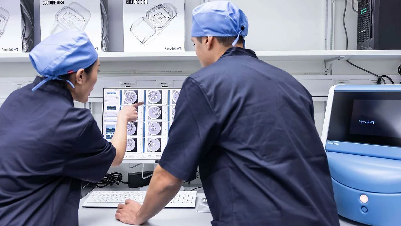
The University of California, San Francisco (UCSF) Centre for Reproductive Health (CRH), one an academic fertility center performing approximately 3,000 IVF cycles annually, has successfully optimized its laboratory operations through the exclusive implementation of EmbryoScope time-lapse technology. This strategic adoption, spearheaded by Mitchell Rosen, MD, and IVF Laboratory Supervisor Philip Marsh, has dramatically improved workflow, enhanced patient care, and addressed critical growth challenges identified by the center in 2017.
Facing constraints such as limited physical space, a shortage of skilled embryologists, and the need to scale operations from 2,000 to 3,000 cycles, UCSF sought a solution to streamline its traditionally time-intensive embryo monitoring processes. Following a comprehensive trial, the EmbryoScope system was fully implemented, allowing continuous monitoring of embryo development without requiring removal from the controlled incubator environment.
The resulting efficiency improvements have been substantial. The elimination of manual handling for assessment has yielded a daily time saving of 7 embryology hours, nearly equivalent to the workload of a full-time embryologist. This efficiency allowed UCSF to restructure its daily operations, successfully transitioning embryo transfers to morning time slots, thereby significantly increasing daily transfer capacity. This shift also provided physicians greater flexibility to perform transfers for their own patients. Furthermore, operational improvements have extended staff benefits, allowing them to complete daily procedures earlier, enhancing work-life balance and job satisfaction.
Beyond workflow, the technology has transformed the precision of embryo evaluation. The time-lapse imaging provides embryologists with critical additional data points to distinguish between similarly graded blastocysts. EmbryoScope enhances the accuracy of procedures like fertilization checks by allowing review of a sequence of images rather than a single time point. Quality and safety measures have also improved dramatically, with reduced foot traffic and dish handling decreasing potential handling errors and environmental exposure (such as fluctuations in pH and temperature).
The continuous monitoring capabilities of the UCSF EmbryoScope are also driving future advancements in reproductive research at UCSF. The large database of developmental patterns being collected is laying the groundwork for the integration of artificial intelligence (AI) in embryo evaluation and selection, positioning UCSF at the forefront of advancing reproductive technology. Through enhanced standardisation, streamlined documentation, and improved transparency facilitated by visual documentation for patients, UCSF demonstrates that the transition to exclusive time-lapse monitoring is a valuable investment that supports growth while maintaining clinical excellence.


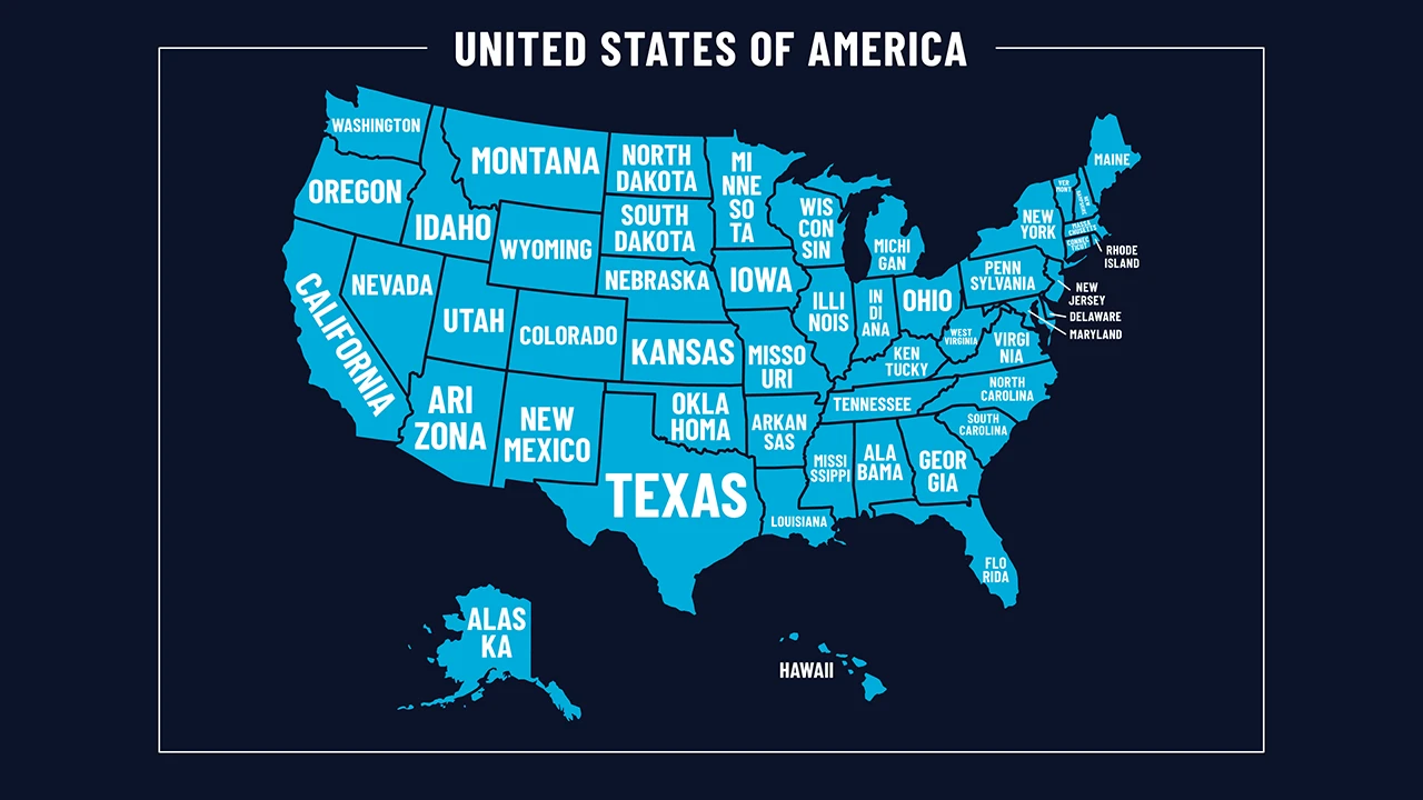
USA Fertility Coverage is expanding in 2026 as more states introduce insurance mandates for IVF, diagnosis, and fertility preservation. While private insurance reforms gain traction, Medicaid coverage remains limited. States are focusing on targeted, incremental changes, creating a varied national landscape with improving but uneven access to reproductive healthcare services.

ASRM analyzes the White House IVF announcement, highlighting drug-pricing deals, new employer benefit pathways, and the unresolved legal and equity questions shaping the future of IVF access in the U.S.
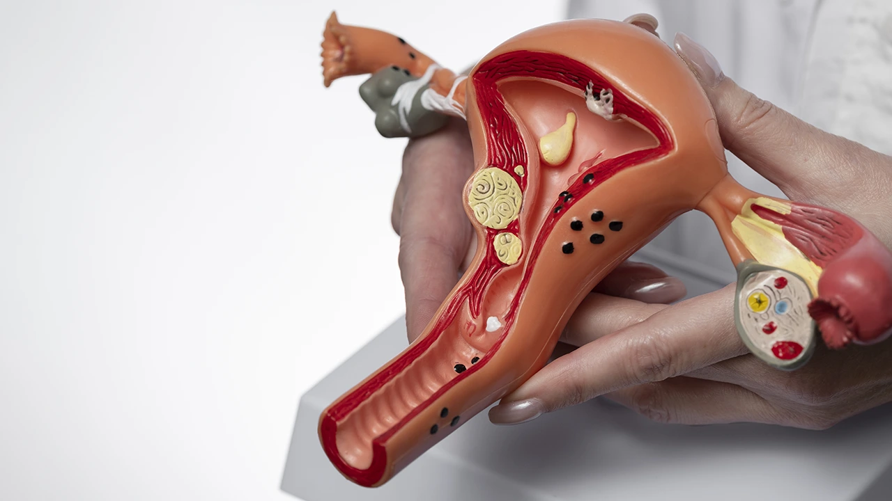
NICE Fertility Guidance has introduced a dedicated pathway for endometriosis, replacing outdated classifications with a personalised approach. The update provides clearer treatment steps, supports healthcare professionals with structured guidance, and aligns with NHS goals to improve access, consistency, and quality of fertility care for patients across the UK.
The National IVF and ICSI Guideline introduces unified clinical protocols for IVF and ICSI in Ireland, strengthening safety measures, promoting eSET, and standardizing assessments and treatment pathways to elevate nationwide fertility care.
CryoFuture’s IVF lab emergency preparedness white paper details key risks to cryogenic storage and offers strategies for monitoring, response and off-site backup to protect reproductive specimens and operations.