Home
 ReportReport
Researchers at the Babraham Institute and Stanford University have engineered a 3D laboratory model of the human uterus to observe embryo implantation in unprecedented detail, up to day 14 of development. Published in Cell, this breakthrough opens new pathways for understanding IVF failure, miscarriage, and pregnancy complications.


For decades, one of the most consequential moments in human reproduction has remained almost entirely out of reach for scientists. Now, a team of researchers from the Babraham Institute and Stanford University has built something unprecedented: a three-dimensional laboratory replica of the womb lining capable of replicating the process by which a human embryo embeds itself into the endometrium. The research, published in the journal Cell, represents one of the most significant advances in reproductive science in recent years, one that could have lasting implications for the millions of people who struggle with infertility and failed IVF cycles.
Approximately one week after fertilisation, the embryo, by this point a compact ball of cells, must successfully adhere to and invade the luminal lining of the uterus to establish a pregnancy. This moment, brief and biologically intricate, has long defied rigorous scientific investigation. The core challenge is observation. There has been no reliable way to replicate the human uterus to observe embryo implantation with the kind of biological accuracy required to understand what is actually happening at the cellular and molecular level.
That barrier has now been cleared.
The 3D model that the team constructed is built around the two primary cell types that make up the endometrium: epithelial cells, which line the surface of the womb, and stromal cells, which form the deeper structural layer beneath them. Both were isolated from endometrial biopsies donated by healthy volunteers. The stromal cells were then embedded into a specially formulated gel, incorporating the same extracellular matrix proteins found in real endometrial tissue, to allow them to grow in a dense, three-dimensional layer. Over this foundation, the epithelial cells were seeded, spreading across the surface to form what became a remarkably accurate replica of the womb lining's architecture.
When cross-sections of the engineered tissue were compared to biopsies of actual human endometrium, the structural resemblance was striking. The model also responded appropriately to hormonal signals, a critical indicator that the engineered womb lining was genuinely receptive to implantation, not merely a visual approximation.
With the model assembled, the research team introduced early-stage human embryos sourced from donated IVF procedures. What they observed were the expected and essential hallmarks of implantation: the embryo adhered to the epithelial surface and began to invade the endometrial scaffold beneath. The process mirrored what is understood to occur in the body, validating the model's fidelity. Crucially, this was not simply an observation exercise. The implanted embryos began secreting human chorionic gonadotropin (hCG) — the hormone detected by pregnancy tests, along with other pregnancy-associated proteins. What made this particularly significant was the response of the engineered womb lining itself.
"We were really excited to see that our system released essential factors that are needed to nourish the embryo in the first few weeks of pregnancy," said Dr. Peter Rugg-Gunn, senior group leader at the Babraham Institute who led the study. "Previous models haven't been able to achieve this, so this represented a breakthrough for us."
This bidirectional response, the embryo signalling to the endometrium and the endometrium signalling back, is precisely what distinguishes a functional pregnancy environment from a passive laboratory construct. Prior systems had been unable to capture it.
The model did not stop at implantation. It supported continued embryo development through to days 12–14 post-fertilisation — a stretch of development that is, in the words of the researchers, "largely unexplored." This is the legal limit for such studies in many jurisdictions, and reaching it marked a milestone that had never before been achieved outside the human body.
During this post-implantation window, the research team observed several important developmental milestones. Specialist cell types appeared within the embryo, and the early precursor structures for the placenta began to form. The structures observed are critical for the eventual maternal–foetal exchange of oxygen and nutrients, the biological infrastructure of a healthy pregnancy.
Using single-cell analysis at the sites of implantation, the researchers were able to profile the molecular dialogue taking place between the embryo and the endometrial model, essentially listening in to a conversation that had, until now, been entirely private. Dr. Irene Zorzan, co-first author of the study and a postdoctoral fellow, described what the model makes possible: "Embryo implantation and post-implantation development are crucial events normally hidden from view, and this has limited our ability to explore the cellular and molecular mechanisms underlying this critical phase. Now, we can witness the unexplored aspects of the earliest moments of development and uncover new insight into how the foundations of a successful pregnancy are laid."
For all its scientific elegance, the research carries an urgency rooted in a stubborn clinical reality. Roughly 75% of embryos transferred during IVF procedures fail to implant, according to a study published in the journal Frontiers in Endocrinology. That failure rate is the central limitation of IVF as a treatment and understanding it has been constrained by the very same observational barriers this model now overcomes.
"Understanding embryo implantation and embryo development just after implantation has significant clinical relevance as these stages are particularly prone to failure," said Dr. Rugg-Gunn. "In particular, the high rate of implantation failure represents one of the main limiting factors for IVF success."
The model also creates a direct pathway to investigating recurrent implantation failure (RIF), a diagnosis given to patients who have experienced more than three unsuccessful IVF and embryo transfer attempts despite the transfer of good-quality embryos. By building personalised endometrial models using tissue from RIF patients, researchers can begin to identify what distinguishes a receptive endometrium from one that consistently fails to accept an embryo.
This is the promise that makes the creation of a workable model of the human uterus to observe embryo implantation so significant, not just for basic science, but for clinical medicine. The IVI Foundation and IIS La Fe in Valencia, Spain – both contributing institutions, have already pointed to the potential to design more personalised therapeutic strategies for patients with fertility challenges, built on the molecular insights this model can now provide.
Beyond diagnostic potential, the research team envisions the model as a tool for testing interventions. Treatments designed to increase endometrial receptivity could be evaluated in the model before being advanced to clinical trials. Conditions such as pre-eclampsia, which has its origins in abnormalities of early placental development, could potentially be modelled and studied in ways that have not previously been possible.
The study was led by Dr. Matteo Molè of Stanford University and Dr. Francisco Domínguez of the IVI Foundation and IIS La Fe, with collaboration from Cambridge University and the Babraham Institute in Cambridge, UK. It was published in Cell following a rigorous peer-review process, a mark of the scientific community's recognition of its significance. Dr. Molè subsequently received $2.3 million in funding from the California Institute for Regenerative Medicine (CIRM) for the follow-on project titled "Dissecting the cellular and molecular interactions established between a human embryo and maternal endometrium at implantation," a proposal that received a top score of 90 in an exceptionally competitive selection process that drew 372 applicants for just 23 funded projects.
Source: https://news.stanford.edu/stories/2026/05/3d-uterus-model-could-embryo-implantation-ivf-infertility


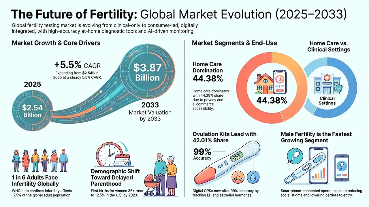
The global Fertility Testing Devices market, valued at USD 2.54 billion in 2025, is projected to reach USD 3.87 billion by 2033, growing at a CAGR of 5.5%. Rising infertility rates, delayed parenthood, and technology-driven at-home testing solutions are fueling robust worldwide demand across North America, Asia Pacific, and beyond.

Global fertility tourism is projected to reach US$13,080.0 Mn by 2032 at a 30.3% CAGR, led by IVF and a strong North America share.
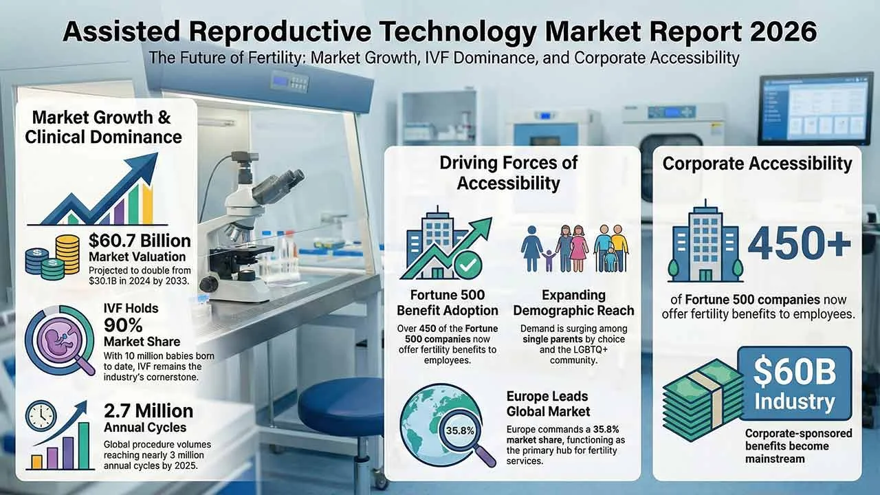
The assisted reproductive technology market continues to expand as infertility rates rise and parenthood is increasingly delayed worldwide. Valued at US$30.1 billion in 2024, the market is projected to reach US$60.7 billion by 2033, supported by IVF innovation, growing awareness, improved accessibility, and sustained investment in reproductive healthcare infrastructure globally.
The global human reproductive technology market is set to reach 45.4 USD billion by 2035, expanding from 27.7 USD billion in 2024 at a 4.6% CAGR. Growth is fueled by technological advancements in fertility treatments, rising infertility rates, delayed pregnancies, and increasing acceptance of single and LGBTQ+ parenting worldwide.
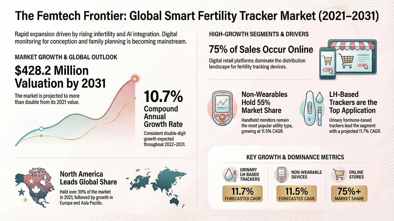
This comprehensive report analyses the Smart Fertility Tracker Market, detailing its projected growth from US$ 160.2 million in 2021 to US$ 428.2 million by 2031. It examines key drivers such as rising infertility and technological advancements in AI-integrated hormone monitoring, alongside detailed segmentation by utility, application, and region.
The Sperm Separation Systems Market is projected to exceed USD 1.5 billion by 2035, driven by automation, microfluidics, and rising male infertility awareness.