Home
 ReportReport
Human preimplantation embryo arrest in ART remains a major challenge in IVF, affecting early embryo development and success rates. Driven by genetic mutations, epigenetic disruptions, and metabolic failures, this condition halts growth before implantation, prompting advances in AI-based embryo selection and precision medicine to improve reproductive outcomes.


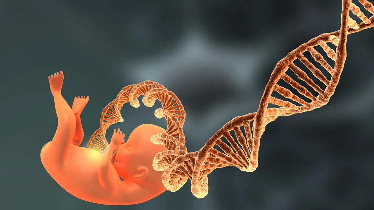
For many individuals and couples, assisted reproductive technology (ART), such as in vitro fertilisation (IVF), represents a beacon of hope for building a family. However, the journey is often marked by a significant biological hurdle known as human preimplantation embryo arrest, or PREMBA. This phenomenon occurs when an embryo, which initially appears to be developing normally in a laboratory setting, suddenly stops growing before it can be transferred to the womb.
In clinical settings, PREMBA is a major challenge, affecting approximately 10% of embryos during the early stages of cell division. Even more striking is that roughly 60% of all embryos created through IVF or intracytoplasmic sperm injection (ICSI) fail to reach the "blastocyst" stage, which is the final stage of development before implantation. This frequent failure leads to a significant emotional and financial burden for patients who face repeated unsuccessful cycles. To address this, scientists are working to uncover the genetic and molecular reasons why some embryos "stall" while others thrive. Currently, researchers categorise these failures into two groups: those caused by known genetic mutations and those that remain "unexplained" but are linked to complex internal biological errors.
To understand human preimplantation embryo arrest in ART, one must first understand the "maternal-to-zygotic transition" (MZT). When an egg is first fertilised, it does not immediately use its own DNA to grow. Instead, it relies on instructions and materials, primarily proteins and molecules called mRNA, inherited from the mother's egg.
As the embryo begins to divide, it must perform a delicate biological baton pass. It needs to "clean up" the mother’s leftover instructions and "awaken" its own genetic blueprint, a process called zygotic genome activation (ZGA). If this handover is disrupted, the embryo enters a state of developmental "stagnation", often stopping at the very moment it was supposed to take control of its own growth.
Scientists have identified 24 specific genes that, when mutated, can cause an embryo to stop developing. These genetic errors are often compared to a "Mendelian disorder", meaning they follow predictable patterns of inheritance. These mutations generally fall into several functional groups that act as the building blocks of early life.
One group involves the "cleanup crew" responsible for removing the mother's genetic leftovers. Genes like BTG4 and ZAR1 are essential for this process. If these genes are faulty, the old maternal instructions clutter the embryo, preventing the new embryonic programme from starting. In some cases, embryos with BTG4 mutations stop growing at the one-cell stage because they simply cannot move past the starting line.
Another critical group is the "internal skeleton" of the cell. For an embryo to divide, it must build a structure called a "spindle" to pull its DNA apart into two new cells. Mutations in genes like TUBB8 and TUBA4A disrupt this internal scaffolding, leading to severe fragmentation or a total stop in division. Interestingly, research has shown that some mutations in a gene called CHEK1 act like a permanent "stop sign" for the cell cycle. In a promising discovery, scientists found that using a specific chemical inhibitor could essentially "turn off" this stop sign, allowing arrested embryos to recover and develop into high-quality blastocysts.
Furthermore, a complex known as the Subcortical Maternal Complex (SCMC) acts as a structural toolkit unique to mammalian eggs. It helps organise the cell and ensures that the early divisions are symmetrical. When genes that form this complex, such as PADI6 or TLE6, are mutated, the embryo may divide unevenly or stop growing between the two-cell and eight-cell stages.
While genetic testing can identify some causes, many cases of human preimplantation embryo arrest in ART occur in embryos that have no obvious genetic mutations. To solve this mystery, researchers use "multi-omics", which is the study of multiple layers of biological information, such as how DNA is packaged, how genes are turned on, and how the cell produces energy.

In these unexplained cases, the embryo often suffers from a "molecular traffic jam". Even if the embryo looks normal under a microscope, its internal "clock" is out of sync. For example, some embryos fail to activate specific "master regulator" genes, such as YY1 or primate-specific genes like DPRX and ARGFX. Without these regulators, the embryo cannot "wake up" its own genome, leading to a complete halt in development.
Beyond the genetic code itself, there is a layer of "chemical tags" on the DNA called epigenetics. These tags, such as DNA methylation, act like volume knobs or light switches, telling the cell which genes to turn on or off. For an embryo to grow, it must "reset" these tags by removing the mother's and father's old patterns.
In many arrested embryos, this reset fails. The DNA remains "hypermethylated", or tightly locked, meaning the embryo cannot access the genes it needs to survive. This often silences essential genes responsible for metabolism, such as the glucose transporter SLC2A3.
Because the embryo cannot process sugar correctly, it experiences a "metabolic collapse". Instead of switching to a high-energy fuel source called glycolysis, which is necessary for the final stages of growth, arrested embryos stay stuck in a low-energy state. This energy shortage eventually pushes the embryo into a "senescent-like" state, which is essentially a form of biological ageing where the cell stops dividing but remains alive in a dormant, stressed condition.
Finally, the physical integrity of the DNA plays a major role. Chromosomal aneuploidy, having the wrong number of chromosomes, is a frequent cause of human preimplantation embryo arrest in ART, particularly in older patients. While some embryos with chromosomal errors can still reach the blastocyst stage, many encounter "chaotic" divisions that are simply incompatible with life. These embryos often have disorganised internal structures and high levels of DNA damage, which trigger a permanent stop in growth to prevent the development of a non-viable pregnancy.
The ultimate goal of studying human preimplantation embryo arrest in ART is to move away from a "trial and error" approach and toward precision diagnosis. By understanding the specific molecular reasons why an embryo stops growing, doctors can develop more targeted strategies to help patients.
One exciting avenue for the future is the integration of artificial intelligence (AI) with time-lapse imaging. By training computers to recognise subtle patterns in how an embryo moves and divides in its first few hours of life, clinicians may be able to predict which embryos are at high risk of arresting before it even happens. This would allow for better embryo selection and potentially higher success rates for IVF treatments.
Additionally, researchers are investigating the use of "rescue" molecules. Treatments like SIRT agonists or specific activators have shown promise in laboratory settings for "unlocking" stagnant epigenetic patterns or boosting an embryo's failing metabolism. While these interventions are still in the research phase, they offer a glimpse into a future where "stalled" embryos could be given the biological jumpstart they need to continue their journey.
Human preimplantation embryo arrest remains a complex and challenging hurdle in the field of reproductive medicine. Whether caused by a specific inherited mutation or a systemic failure of the embryo's internal machinery, the result is a heartbreaking stop in potential life. However, by bridging the gap between genetics, metabolism, and advanced technology, scientists are slowly unravelling the "symphony" of molecular events required for a single cell to become a human being. These discoveries not only deepen our understanding of early human life but also pave the way for more effective, personalised fertility treatments that can turn the dream of parenthood into a reality.


Researchers at the Babraham Institute and Stanford University have engineered a 3D laboratory model of the human uterus to observe embryo implantation in unprecedented detail, up to day 14 of development. Published in Cell, this breakthrough opens new pathways for understanding IVF failure, miscarriage, and pregnancy complications.

Global fertility tourism is projected to reach US$13,080.0 Mn by 2032 at a 30.3% CAGR, led by IVF and a strong North America share.
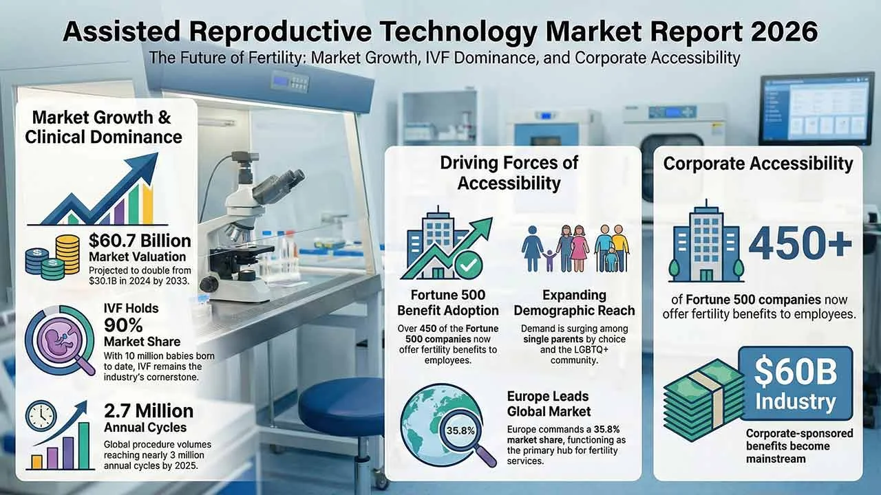
The assisted reproductive technology market continues to expand as infertility rates rise and parenthood is increasingly delayed worldwide. Valued at US$30.1 billion in 2024, the market is projected to reach US$60.7 billion by 2033, supported by IVF innovation, growing awareness, improved accessibility, and sustained investment in reproductive healthcare infrastructure globally.
The global human reproductive technology market is set to reach 45.4 USD billion by 2035, expanding from 27.7 USD billion in 2024 at a 4.6% CAGR. Growth is fueled by technological advancements in fertility treatments, rising infertility rates, delayed pregnancies, and increasing acceptance of single and LGBTQ+ parenting worldwide.
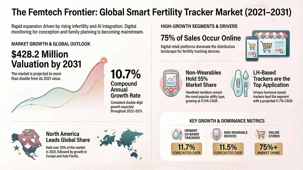
This comprehensive report analyses the Smart Fertility Tracker Market, detailing its projected growth from US$ 160.2 million in 2021 to US$ 428.2 million by 2031. It examines key drivers such as rising infertility and technological advancements in AI-integrated hormone monitoring, alongside detailed segmentation by utility, application, and region.
The Sperm Separation Systems Market is projected to exceed USD 1.5 billion by 2035, driven by automation, microfluidics, and rising male infertility awareness.

The Human Reproductive Technologies market is poised for steady growth through 2032, driven by strategic moves from leading players, segmentation expansion, and region-specific demand shifts.