Home
 NewsNews
Scientists capture the first real-time 3D images of human embryo implantation, revealing dynamics that could transform infertility and IVF treatment.


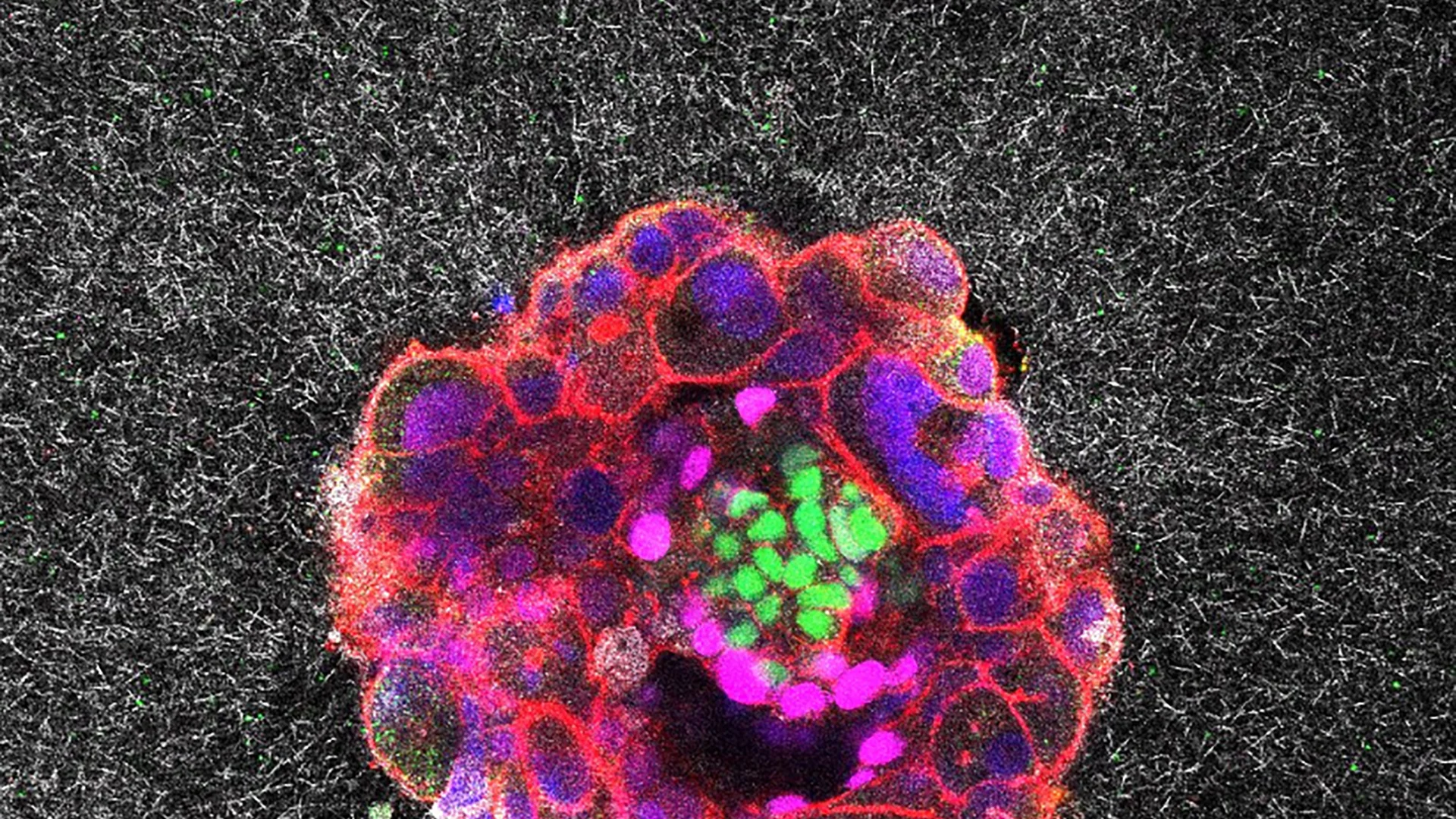
Researchers have, for the first time, captured real-time, three-dimensional images and videos of a human embryo implanting into tissue designed to mimic the human uterine environment. This groundbreaking research, largely carried out at the Institute for Bioengineering of Catalonia (IBEC) in collaboration with Dexeus University Hospital in Barcelona, has given scientists unprecedented visual access to the earliest stages of human pregnancy, offering new insight into one of medicine’s biggest mysteries: why some embryos fail to implant, causing infertility or early miscarriage.
Before this study, scientists relied on static snapshots of embryo implantation, leaving much of the process a mystery. Implantation is the pivotal event when a fertilized embryo attaches and integrates into the uterine lining, a stage responsible for about 60% of miscarriages and a leading cause of infertility. Using an innovative 3D gel platform based on collagen (a key uterine component), the IBEC team successfully visualized human embryos as they burrowed into, and radically reorganized, the uterine matrix.
Implantation failure is a dominant barrier in both natural and assisted reproduction, including in vitro fertilization (IVF). The new imaging system allows scientists and clinicians to monitor how embryos interact with a simulated uterine matrix, offering avenues to:
The researchers found that human embryos exert significant force as they burrow into the uterine tissue. This process is not passive; it’s “surprisingly invasive,” as the embryo pushes into, pulls on, and merges with its environment. The forces are vital for the embryo to invade the uterine tissue and become completely integrated, an essential step to begin a pregnancy. This intense level of activity helps explain why some women experience cramps or mild bleeding at the time of implantation.
Observing this invasive process in real time has not previously been possible, as the action takes place entirely inside the woman’s body and is shielded from direct observation. According to Samuel Ojosnegros, the lead researcher, “What happens between the transfer and the first ultrasound weeks later is a black box.” Implantation failure during this stage is responsible for approximately 60% of miscarriages and is a significant hurdle both for natural conception and in assisted reproduction like in vitro fertilization (IVF).
One of the most promising outcomes from this research lies in its potential to improve fertility treatments. Implantation remains the least understood and most failure-prone stage of both natural conception and IVF attempts. For IVF, embryos are typically transferred into the uterus five days after artificial fertilization. The events that follow, up to the first detectable pregnancy, have been largely mysterious until now.
With 3D and real-time imaging, scientists can objectively study how embryos interact with uterine tissue, both in terms of chemical signaling (like the release of enzymes that break down surrounding tissue) and biomechanics (the force exerted by the embryo). These insights can guide fertility doctors in optimizing the timing, environment, and methods for embryo transfer, potentially increasing success rates for IVF, which currently remain under 50% per cycle for most patients.
The researchers hope to use these findings to refine assisted reproduction practices and even develop treatments specifically targeting the implantation phase. By understanding what successful implantation looks like in detail, including the movements, forces, and cellular changes, clinicians can better identify what might be going wrong in cases of repeated failure and intervene more effectively.
Key technical and biological insights from species differences to mechanosensitivity underscore just how dynamic and orchestrated implantation truly is. For fertility medicine, this marks a giant step towards more personalized, evidence-based care.


Abu Dhabi University and FertiClinic Group have joined forces to establish the FertiClinic Reproductive Research Lab, an advanced reproductive research lab in Abu Dhabi housing IVF, andrology, and cryopreservation facilities. The partnership supports UAE national priorities, bridges academic and clinical expertise, and advances fertility innovation across the region.
NHS Kent and Medway has overhauled its fertility funding policy in a move that sees NHS cuts IVF access for thousands of patients. Effective 1 April 2026, the eligible age ceiling drops from 40 to 38, funded cycles fall from two to one, and embryo transfers are capped at two.
ASEAN's First Telemedicine IVF by Inspire IVF introduces a new era of remote fertility care. Combining AI technology with expert consultation, the platform enables global access, real-time monitoring, and personalised treatment planning, positioning Thailand as a leading hub for fertility services and advancing digital transformation in reproductive healthcare.
CSG.BIO has gone ahead with the acquisition of Hanabusa IVF, Asian Egg Bank, thereby making it a world leader in reproductive medicine and cutting-edge clinics with enhanced patient reach and advanced laboratory capabilities.
Wisconsin lawmakers from both parties are proposing bills to reduce IVF treatment costs. Republicans suggest a $5,000 tax credit for expenses, while Democrats want mandatory insurance coverage. With IVF cycles costing $20,000-$30,000 and 170,000 residents facing infertility, both parties seek affordable solutions through different approaches.
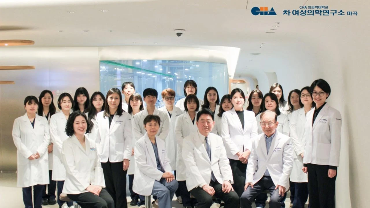
The AI-specialized Infertility Center, which is the largest in Asia, is open in Magok, Seoul and has begun its full-scale medical services from November 17 thereby marking the new era when it comes to fertility in the region.

Alife Health secured CE Mark approval for its AI-powered embryo selection tool, Embryo Predict, under EU MDR, expanding its fertility technology presence across Europe.